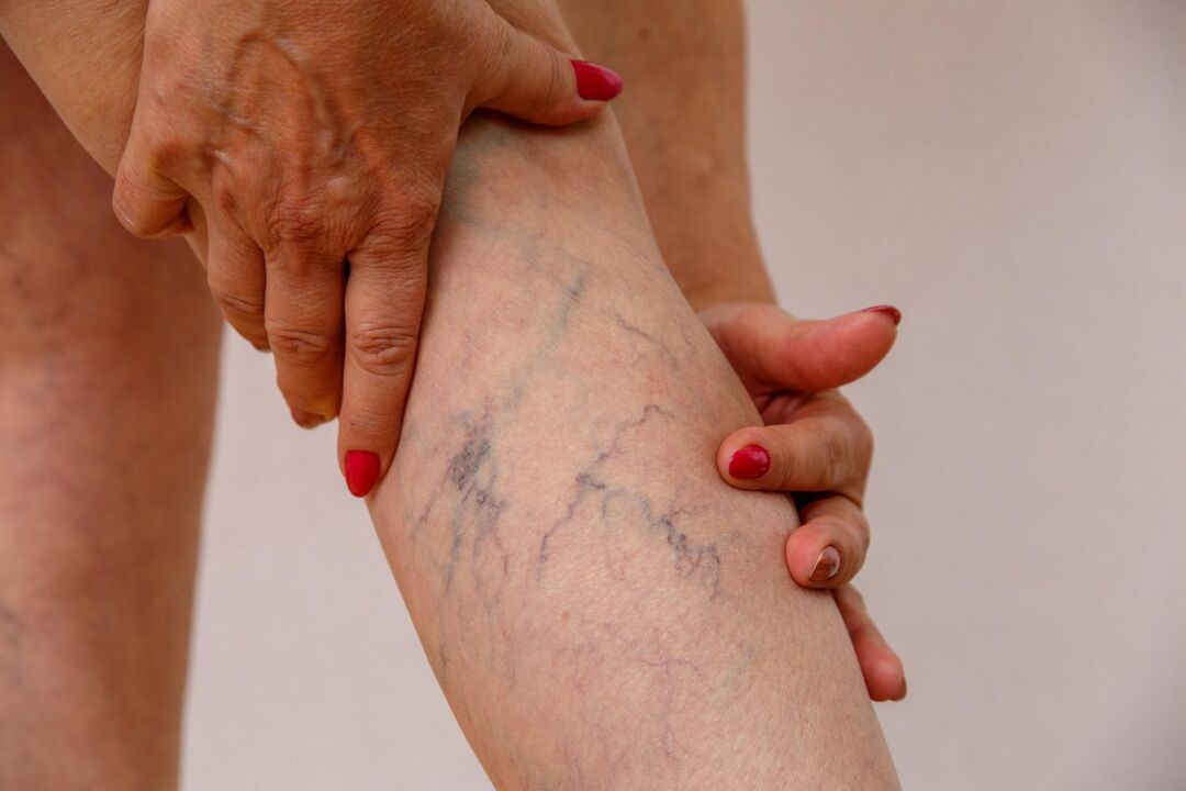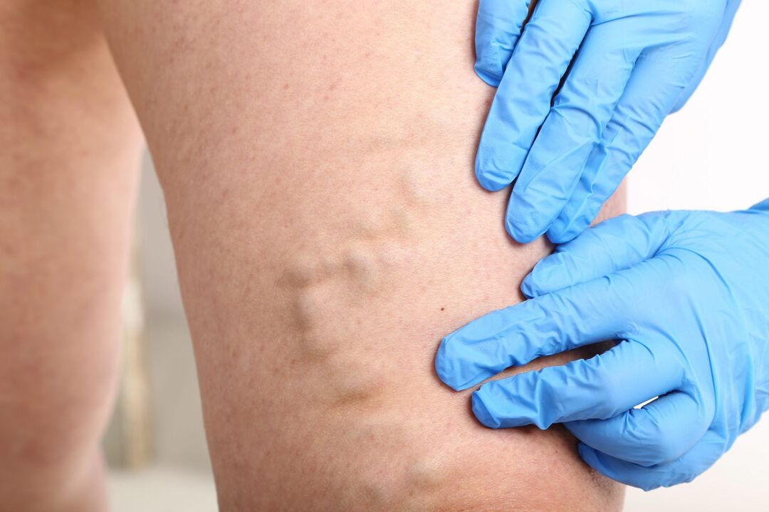
Varicose veins are vascular lesions when the walls of veins stretch against a background of weak connective tissue. The diameter of the vein increases and its wall becomes thinner.
The large diameter of the veins leads to reduced blood flow, congestion of the veins, and causes pain in the lower legs. In this context, varicose veins can lead to thrombophlebitis—inflammation of the affected veins, which is dire for the development of thromboembolic complications. The outer cones visible along the blood vessels allow you to identify varicose veins in the legs. Varicose veins of the lower extremities (ICD code I83) are a very visible condition that can be easily removed.
Esophageal varices are included in the symptoms of portal hypertension, and secondary varices of the perineum in women indicate small pelvic varices and difficulty in outflow of blood from the main veins.
A varicocele (varicocele) presents as secondary pelvic venous hypertension, which can lead to male infertility. The etiology and pathogenesis of varicose veins are very diverse, depending on the localization of the process. An increase in vein diameter is not inherently dangerous, but complications from varicose veins can pose significant health risks and sometimes even life-threatening patients. The causes of leg varicose vein attacks may be strenuous physical exertion, childbirth, and the patient's sedentary lifestyle.
To get an idea of what varicose veins look like, head to Summer Beach. While many varicose vein carriers are embarrassed to be there, you're bound to see how varicose veins manifest in both men and women. This disease is so common, you are sure to see it. After reading this article, you will understand how easy it is to treat leg varicose veins. Don't be afraid to see a phlebologist.
Can we reverse varicose veins?
Many people ask this question in the hope that varicose veins can be cured in the early stages with the help of medication or traditional medicine. If we're talking about varicose veins in the legs, then a phlebologist can definitively answer that question - the degenerative damage to the vein wall doesn't go away without closing the affected vein from the bloodstream or removing it.
A vein that happens to be dilated may still not lose its function and increase in volume due to overflowing blood from the covering part, and the muscle pump in the calf helps blood flow into the deep veins.
Depending on the stage of varicose veins, various surgical and conservative treatments can be applied to stop the progression of varicose veins at different stages. The sequence here is this: if the vein is irreversibly affected, it must be removed or coagulated or glued.
Why are even the initial varicose veins irreversible without surgical intervention? In order to effectively treat leg varicose veins, it is necessary to identify where the pathological secretion of venous blood comes from and remove it with minimal trauma. However, if the phlebologist eliminates the pathological secretions of the veins that cause the varicose and irreversible changes, the enlarged varicose tributaries can restore their function on their own without the need for surgical intervention.
Since varicose vein surgery was first performed on both men and women in the 19th century, modern varicose vein treatment has advanced significantly. According to the degree of varicose veins, the classification of the disease and the appropriate treatment method are compiled.
The clinics of the Innovative Vascular Center know how to treat varicose veins with minimal medical, psychological and cosmetic inconvenience. We do not need to remove varicose veins according to the classic protocol. In the phlebologist's arsenal, the hemodynamic concept of treating the primary cause of varicose veins involves correcting only pathologically altered venous outflow and removing only the affected veins.
Treatment cannot target the cause of the disease, but the pathogenesis of the problem is known, so treatment can be stopped. In women, the presence of varicose veins on the legs can be an annoying symptom due to aesthetic concerns, but fair sex isn't ready to change the ugly appearance of subcutaneous varicose veins that go unnoticed with large scars. Therefore, the clinic offers cosmetic and radical treatments with the best patient reviews.
some anatomy and physiology

Varicose veins are defined as the primary dilation of the subcutaneous venous trunk of the lower extremities due to congenital, contributing, and producing factors. 40% of adults on earth have a chance of developing varicose veins. In developed countries, signs of varicose veins are detected in half of the population.
The great saphenous vein of the leg is represented by two large venous systems - the great saphenous system and the lesser saphenous system. The saphenous vein originates in the foot, extends along the inner surface of the lower leg to the inguinal area, and flows from the medial side of the common femoral artery into the deep vein of the thigh.
On the way from the trunk and tributaries of the great saphenous vein, short vein trunks can be identified - the perforators that connect it with the deep veins of the calf and thigh, which cause varicose veins away from the trunk. These perforators are designed to facilitate the entry of blood into the deep venous system.
The small saphenous vein forms at the lateral malleolus and is characterized by multiple curves along the posterior surface of the lower leg, where it joins the popliteal vein. Between them, the greater and lesser saphenous veins are connected by separate overflows. There are many venous valves in the subcutaneous trunk that ensure blood flow to the heart and prevent the backflow of blood.
Due to the congenital weakness of the vein wall and the load on it, the endovenous valve device malfunctions and blood begins to move in the opposite direction, causing the saphenous vein to overflow, further stretching and developing severe varicose veins. Therefore, it is impossible to achieve the goal of curing chronic varicose veins without eliminating the pathological secretions of the blood.
The classification of subcutaneous varicose veins of the legs is formed from the name of the disease and the cause of its development, the affected venous pool and the stage of chronic venous insufficiency. Varicose veins of the lower extremities are formed by the following factors:
- Congenital dilatation and weakness of the venous wall and increased venous pressure.
- Venous pressure increases due to prolonged lifestyle, strenuous physical exertion, pregnancy and childbirth.
- Congenital and acquired obstruction of venous outflow (compression syndrome, tumor and bone formation compressing the vein).
- Sequelae of previous deep vein thrombosis

Principles of modern varicose vein treatment
The question that many patients often ask is what treatment is needed for varicose veins, if only at the first signs. Varicose veins in the legs are an ongoing and complication-prone condition, so recovery cannot be expected without medical intervention. Consider the primary indication for the treatment of leg varicose veins.
Relieve symptoms of chronic venous insufficiency
Venous hypertension is a subjectively unpleasant consequence of impaired venous outflow, but varicose veins themselves are not harmful. Symptoms of varicose veins that need to be prevented and treated include a feeling of heaviness in the legs, nighttime swelling, increased leg fatigue, and even calf muscle pain. As the disease progresses, venous perforators and deep veins develop stagnation, which can lead to hyperpigmentation of the skin, leading to varicose eczema, and noting heaviness in the lower legs.
The most popular and advertised way to treat the symptoms of varicose veins in the legs is to take various varicose vein pills, use ointments and creams, which makes contacting a specialist late. It is important to understand that these remedies do not affect the course of varicose veins, which is why they only slightly relieve complaints and symptoms in the early stages. The fact that varicose veins go away after treatment with such drugs is not to be expected.
Treatment of varicose vein complications (trophic ulcers, thrombophlebitis, venous bleeding)
In approximately 50% of cases, varicose disease is complicated by a localized inflammatory process, which expands the indications for aggressive surgical strategies. In most cases, patients are treated for varicose-thrombotic phlebitis (ICD code I80) when complications arise, which can cause significant damage or develop trophic ulcers. Occasionally disturbed by nighttime cramping, redness, and pain in the calf muscles.
Treatment of thrombophlebitis can be done conservatively (heparin ointment, lyoton, compression) or more aggressively - removal of the affected varicose veins or its laser coagulation. Clinical recommendations do not give a definitive answer to this question, but with a positive approach, in addition to thrombophlebitis, its cause is also eliminated, which is varicose veins.
Nutrient ulcer is an extreme manifestation of chronic venous insufficiency and has great danger. It looks like a skin defect in the medial malleolus area with active purulent discharge, loose granulation, and ongoing damage to the surrounding subcutaneous tissue.
A varicose ulcer that begins is prone to progression and responds very poorly to conservative treatment. The best treatment available today is laser correction of the venous outflow tract (EVLK) of the great or small saphenous varices and proper topical treatment (special dressings, cleaning of the ulcer). One does not work without the other, so there is no need to rely solely on ointments to heal trophic ulcers. A mandatory component of treatment is compression therapy with the help of special compression stockings. The patient's complaints were greatly alleviated.
Cosmetic indications for varicose veins
Varicose veins are a condition that rarely leads to dangerous complications, but usually requires you to turn to a specialist. Swollen varicose veins cause a lot of aesthetic problems for their owners. Often younger patients are embarrassed by these knots and hide their legs. If men are less afraid of varicose veins and they can walk in pants more often, women will always want to walk with open legs.
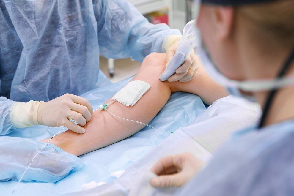
The good news is that advanced varicose veins in the legs of women or men can now be removed with a single laser photocoagulation procedure without leaving any traces. Modern interventions are performed with minimal puncture, no incision required, and are completely invisible 3-4 weeks after the intervention. The patient is brought to the operating table under local anesthesia, and the procedure lasts 40-50 minutes. Lasers have amazing cosmetic results and stable recovery from varicose manifestations, which is why EVLT is popular among physicians and younger patients with leg varicose veins of any stage.
Prevent the development of varicose vein complications
The solutions to these problems can be solved by conservative and actionable methods. The main goal of modern phlebology is to minimize the surgical trauma of treating varicose veins and to have the longest possible therapeutic and cosmetic results. In order to solve the first problem, it is necessary to block the reversely working venous vessels through which significant discharge occurs, and to solve the second problem, the dilated veins need to be removed or closed from the blood circulation.
Diagnosis of varicose veins
To properly diagnose superficial venous disease, an examination by an experienced specialist and an ultrasound scan of the great saphenous and deep veins from the abdomen to the feet are required. Information from these study methods was sufficient to correctly identify the diagnosis in the vast majority of patients. The main signs of varicose veins in the legs can be identified with the naked eye, and an ultrasound can be used to determine the cause.
In some cases, doctors do an invasive test of the amount of venography on an angiography device. After treatment, the patient needs to regularly monitor the condition of the surgical vein, which doctors use to monitor using diagnostic ultrasound. If physicians have doubts about the status of the deep veins at the diagnostic stage, diagnostic MRI or contrast CT can accurately determine their patency.
Treatment of central varicose veins
Vascular surgeons can only cure varicose veins of the lower extremities by eliminating the cause of their appearance. It is necessary to fight the causes of the development of varicose veins and the progression of the disease. Consider major techniques that have proven effective.
Varicose Vein Laser Therapy (EVLT)
Intravenous laser coagulation is based on heating the vein wall with a coherent beam. Varicose veins can be effectively treated without incisions and general anesthesia. The fiber optic is inserted into the vein by puncture under ultrasound guidance. The laser energy of a certain wavelength is absorbed by the vein wall at the moment of its occurrence, causing its heating and the destruction of the connective tissue. As a result, the walls of the veins become scar tissue, and blood flow to the affected veins stops completely. Achieves the same effect as surgical removal of a vein, but without the incision, general anesthesia, and pain.
In terms of its effectiveness, EVLK surpasses open surgery for phlebotomy. Regardless of the degree of lymph node development, 98% of surgical patients recover from varicose veins. Rare side effects include skin numbness in the coagulated vein area, inflammation and blood clots in the coagulated vein. The overall incidence of such complications does not exceed 1%. At Innovative Vascular Center, EVLK is the "gold standard" that cures any early and late varicose veins. Patients leave the best reviews after laser treatment.
Varicose Vein Radiofrequency Occlusion (RFO)
In terms of its impact and effectiveness, RFO, like laser, is known as a thermal treatment for varicose veins, but uses different physical principles. Radiosondes are also inserted into veins by puncture. The intervention is performed under local anesthesia. The RFO principle is based on thermal energy generated in the probe, which is then transferred to the vessel wall. Heating the wall causes thermal destruction of its structural elements, followed by scarring of the veins.
Both methods (EVLK and RFA) involve thermal ablation (thermal) techniques. They are similar in terms of their effectiveness, however, the laser heats the vein wall itself, while the RFO heats the working surface of the probe, and heat is transferred to the wall through the liquid portion of the blood.
According to experts, EVLT more completely destroys the structure of the affected veins, therefore, recurrence after laser is less frequent than radiofrequency occlusion. Doctors noted that 98% of varicose veins did not recur after EVLK and 86% after RFO. Based on 20 years of experience, phlebologists have concluded that heat therapy for varicose veins is more effective than traditional phlebotomy.
Athermal methods of occluding varicose veins
In the 1970s, surgeons began to show increasing interest in minimally invasive surgical treatment of varicose veins and began using electrocoagulators. Good idea, but poorly implemented in practice. The patient had skin burns, which may be why doctors were afraid to use hyperthermia for varicose veins long-term. Chemical methods for venous occlusion have been shown to be safe and very effective. These include various variants of sclerotherapy and adhesive removal.
Sclerotherapy
Sclerotherapy is the intravenous administration of special drugs that cause damage to the vein walls and then occlude (overgrow) the lumen of the varicose veins. The history of this method begins in the 19th century and has an interesting development path. At the Vascular Center, specialists use state-of-the-art technology - sclerotherapy in the form of a foam. Treatment that lasts six months can keep you free of varicose veins from your lower extremities for a long time. Although the recurrence rate within 5 years is about 50%. With sclerotherapy, the treatment does not focus entirely on the cause of the varicose veins, but rather eliminates the venous knot itself, so it can be combined with other minimally invasive methods (EVLK, RFO). A feature of sclerotherapy is the appearance of dense cone-coagulation at the site of sclerotic veins, which resolves for up to six months.
Glue the varicose veins with special glue
The Venaseal technique is the name given to a method of athermally occluding a varicose vein of the great saphenous vein, which involves the introduction of a special glue into the lumen of the vein, which polymerizes within the lumen of the vein, causing it to become blocked. The idea looks interesting and has evolved over the past decade, but it has some flaws. First, the glue remains in the affected vein as a foreign body and does not dissolve. Second, there is a risk of phlebitis around closed veins due to the body's reaction to the foreign body. Third, it is an expensive treatment.
Treating varicose veins with this method costs about twice as much as laser photocoagulation. There are no long-term studies on the long-term outcomes of this treatment. The advantages of this technology have not yet been established, but research is actively underway, and varicose veins have the potential to become a disease in which the entire treatment regimen becomes a "magic" injection. It is characterised by the fact that this method has not been considered in the latest clinical guidelines, but is actively offered by some phlebology centres.
Surgical treatment of lower extremity varicose veins
Since the mid-19th century, doctors have been dealing with how to get rid of large varicose veins in the superficial veins of the legs and prevent complications. The history of battling enlarged veins clearly shows that from the early days of large incisions deforming the legs, surgery has progressed to micropuncture, which allows you to manage varicose veins without cosmetic defects.
Advanced phlebologists use elements of classic surgery to remove individual varicose veins and tributaries using puncture in the form of venuleectomy. This is probably the most aesthetically pleasing way to remove thin-skinned varicose veins. One month after such an operation, the skin was not even red.
Other thermal methods
Phlebologists often use bizarre methods when deciding how to treat varicose veins. Varicose veins are treated with superheated steam and bipolar coagulation. However, modern hyperthermia methods are more effective, they allow doctors to prevent further development of varicose veins, and patients can be treated on an outpatient basis without affecting their lifestyle. In the hands of a novice phlebologist, thermal ablation methods can lead to unpleasant complications: reduced sensitivity, burns, sealing. This method is more than 98% effective in the hands of an experienced phlebologist, and the laser method and RFO not only get you out of the original form, but also get rid of severely visible varicose veins in the legs without incisions.
Use special glue
This method has attracted great interest among phlebologists since its inception. It involves gluing the trunk of the great saphenous vein with special cyanoacrylate glue. In the lumen of the blood vessel, this glue polymerizes and fills the lumen of the dilated blood vessel. As the developers envisioned, this method does not require any anesthesia, and a "plug" appears in the blood vessel, which reliably blocks blood flow. Given this, half an hour is enough to get rid of varicose veins in the legs. Venasil is the only technology that can treat varicose veins without wearing compression stockings.
Most women can resume normal activities immediately. Symptoms of chronic venous insufficiency resolve quickly after surgery. The process of aggressive promotion of this glue on the intravenous market should begin in the near future. However, there are also certain disadvantages: There are foreign bodies in the human body. The crimped glue can remain in the blood vessels forever and can lead to chronic allergies, sometimes with inflammation of the vessel wall or purulent rejection of the polymer. Acute thrombophlebitis of adherent vessels may occur.
Using glue on the trunk of the saphenous vein does not eliminate the need to eliminate tributary varices, which is why doctors must remove signs of subcutaneous varicose veins with sclerotherapy or phlebectomy. The visible effects of using glue can only be seen in combination with other methods of eliminating varicose veins. Patients have to pay more. The unreasonable cost of gluing kits makes the process much more expensive than modern laser or radio frequency methods.
Clinically, the thermal method is preferred. Phlebologists believe that good local anesthesia is better than expensive and untested treatments for leg varicose veins. Also, the results are the same at best. If a recurrence occurs, the patient will have to perform complex procedures to remove the sealed blood vessel, as other methods will no longer work.
Modern approaches to combined treatment of reflux along the subcutaneous venous trunk add extra weight over traditional sclerotherapy. The mechanochemical procedure is understood as a combination of mechanical damage to the inner surface of the vein wall and introduction of a sclerosing agent. The catheter was inserted into the main saphenous vein by puncture under ultrasound guidance. After installing the conduit in the correct location, connect the equipment. The rotating sharp head of the catheter rotates at up to 3500 revolutions per minute, causing significant damage to the lining of the vein wall. At the same time, the sclerosing agent is injected through the catheter, which "mixes" in the lumen of the vessel and acts on the vessel wall with the help of the rotating part of the catheter, causing it to become inflamed and adherent.
This is a modern microsurgical cosmetic method used to remove the tributaries of varicose veins. It means a delicate technique of puncturing and pulling varicose veins with the help of special tools. This operation is not suitable for novice phlebologists, you need to master the skills of fine operation. Veneectomy is a surgical procedure that does not use a scalpel and is performed under local anesthesia. The piercings are done in the direction of the skin line, so they are barely visible after 2 months.


